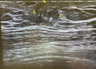
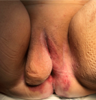
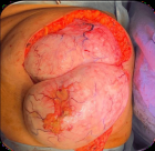
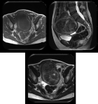
Figure 5
Fibrothecal Tumors of the Ovary - Case Report
M Lamrani*, K Lakhdar, S Sardaoui, Y Alami, F Tijami, H Hachi, Z El-Hanchi and A Baydada
Published: 11 November, 2024 | Volume 7 - Issue 4 | Pages: 117-119
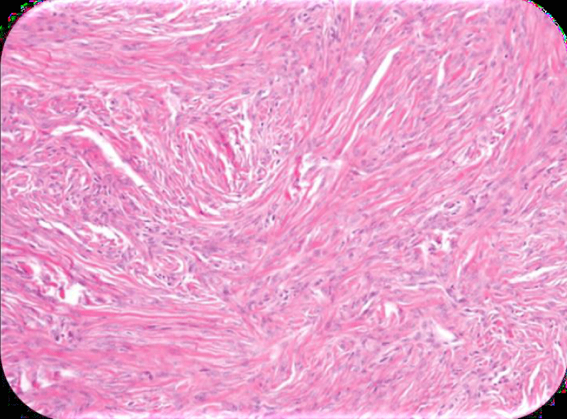
Figure 5:
Figure showing the anatomopathological appearance of an ovarian fibrothecoma on slide. A Hematoxylin and Eosin (H and E) stained slide shows bland spindle cells arranged in fascicles and set within a collagenous stroma [8].
Read Full Article HTML DOI: 10.29328/journal.cjog.1001177 Cite this Article Read Full Article PDF
More Images
Similar Articles
-
Cervical choriocarcinoma in a post-menopause woman: Case report and review of literatureZeinab Nazari,Leila Mortazavi*,Noushin Gordani. Cervical choriocarcinoma in a post-menopause woman: Case report and review of literature. . 2022 doi: 10.29328/journal.cjog.1001101; 5: 019-021
-
Fibrothecal Tumors of the Ovary - Case ReportM Lamrani*,K Lakhdar,S Sardaoui,Y Alami,F Tijami,H Hachi,Z El-Hanchi,A Baydada. Fibrothecal Tumors of the Ovary - Case Report. . 2024 doi: 10.29328/journal.cjog.1001177; 7: 117-119
Recently Viewed
-
Assessment and treatment of patients with kinesiophobia: A Delphi consensusMattias Santi*,Ina Diener,Rob Oostendorp. Assessment and treatment of patients with kinesiophobia: A Delphi consensus. J Nov Physiother Rehabil. 2022: doi: 10.29328/journal.jnpr.1001047; 6: 023-028
-
Sinonasal Myxoma Extending into the Orbit in a 4-Year Old: A Case PresentationJulian A Purrinos*, Ramzi Younis. Sinonasal Myxoma Extending into the Orbit in a 4-Year Old: A Case Presentation. Arch Case Rep. 2024: doi: 10.29328/journal.acr.1001099; 8: 075-077
-
Timing of cardiac surgery and other intervention among children with congenital heart disease: A review articleChinawa JM*,Adiele KD,Ujunwa FA,Onukwuli VO,Arodiwe I,Chinawa AT,Obidike EO,Chukwu BF. Timing of cardiac surgery and other intervention among children with congenital heart disease: A review article. J Cardiol Cardiovasc Med. 2019: doi: 10.29328/journal.jccm.1001047; 4: 094-099
-
Advancing Forensic Approaches to Human Trafficking: The Role of Dental IdentificationAiswarya GR*. Advancing Forensic Approaches to Human Trafficking: The Role of Dental Identification. J Forensic Sci Res. 2025: doi: 10.29328/journal.jfsr.1001076; 9: 025-028
-
Scientific Analysis of Eucharistic Miracles: Importance of a Standardization in EvaluationKelly Kearse*,Frank Ligaj. Scientific Analysis of Eucharistic Miracles: Importance of a Standardization in Evaluation. J Forensic Sci Res. 2024: doi: 10.29328/journal.jfsr.1001068; 8: 078-088
Most Viewed
-
Evaluation of Biostimulants Based on Recovered Protein Hydrolysates from Animal By-products as Plant Growth EnhancersH Pérez-Aguilar*, M Lacruz-Asaro, F Arán-Ais. Evaluation of Biostimulants Based on Recovered Protein Hydrolysates from Animal By-products as Plant Growth Enhancers. J Plant Sci Phytopathol. 2023 doi: 10.29328/journal.jpsp.1001104; 7: 042-047
-
Sinonasal Myxoma Extending into the Orbit in a 4-Year Old: A Case PresentationJulian A Purrinos*, Ramzi Younis. Sinonasal Myxoma Extending into the Orbit in a 4-Year Old: A Case Presentation. Arch Case Rep. 2024 doi: 10.29328/journal.acr.1001099; 8: 075-077
-
Feasibility study of magnetic sensing for detecting single-neuron action potentialsDenis Tonini,Kai Wu,Renata Saha,Jian-Ping Wang*. Feasibility study of magnetic sensing for detecting single-neuron action potentials. Ann Biomed Sci Eng. 2022 doi: 10.29328/journal.abse.1001018; 6: 019-029
-
Pediatric Dysgerminoma: Unveiling a Rare Ovarian TumorFaten Limaiem*, Khalil Saffar, Ahmed Halouani. Pediatric Dysgerminoma: Unveiling a Rare Ovarian Tumor. Arch Case Rep. 2024 doi: 10.29328/journal.acr.1001087; 8: 010-013
-
Physical activity can change the physiological and psychological circumstances during COVID-19 pandemic: A narrative reviewKhashayar Maroufi*. Physical activity can change the physiological and psychological circumstances during COVID-19 pandemic: A narrative review. J Sports Med Ther. 2021 doi: 10.29328/journal.jsmt.1001051; 6: 001-007

HSPI: We're glad you're here. Please click "create a new Query" if you are a new visitor to our website and need further information from us.
If you are already a member of our network and need to keep track of any developments regarding a question you have already submitted, click "take me to my Query."


