Figure 1
Rejuvenation of Ovarian Function after Autologous Platelet Lysate Injection: Promising Evidence from Confirmed Cases
Athanasios Garavelas, Efstathios Michalopoulos*, Panagiotis Mallis and Eros Nikitos
Published: 13 December, 2023 | Volume 6 - Issue 4 | Pages: 225-232
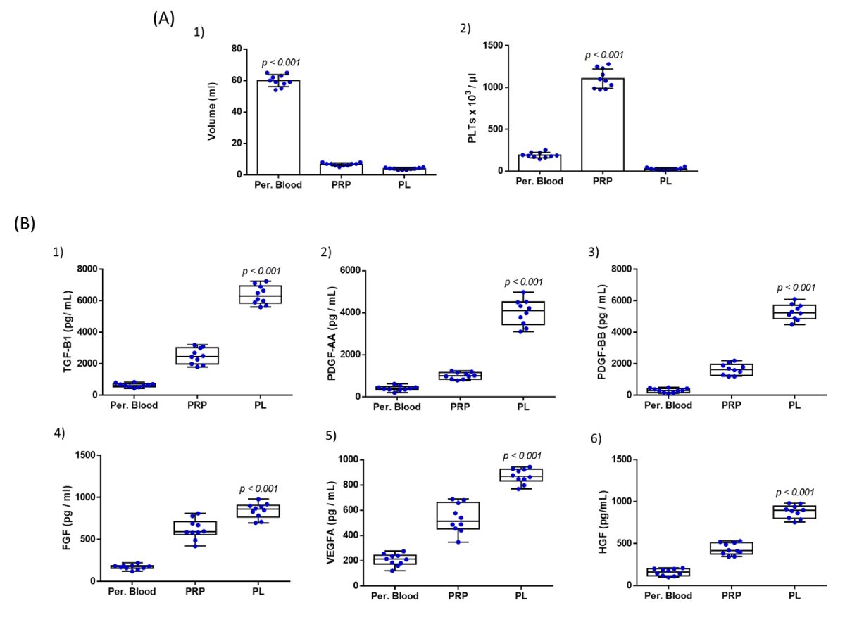
Figure 1:
Figure 1: Quality characteristics of PL involved the determination of volume and PLT count (A) and the biomolecule content quantification (B), in peripheral blood, PRP, and PL samples. Volume determination (A1) and PLT count in peripheral blood (A2), PRP, and PL samples from all participants. Statistically, significant differences were observed between the volume and PLT concentrations between peripheral blood, PRP, and PL samples (p < 0.001). The quantification involved the determination of TGF-β1 (B1), PDGF-AA (B2), PDGF-BB (B3), FGF (B4), VEGF-A (B5), and HGF (B6). Kruskall-Wallis test indicated statistically significant differences between the peripheral blood, PRP, and PL with respect to TGF-B1 (p < 0.001), PDGF-AA (p < 0.001), PDGF-BB (p < 0.001), FGF (p < 0.001), VEGF-A (p < 0.001) and HGF (p < 0.001).
Read Full Article HTML DOI: 10.29328/journal.cjog.1001153 Cite this Article Read Full Article PDF
More Images
Similar Articles
-
TMD and pregnancy?Afa Bayramova*. TMD and pregnancy?. . 2018 doi: 10.29328/journal.cjog.1001001; 1: 001-006
-
Screening of Gestational diabetes mellitusGehan Farid*,Sarah Rabie Ali*,Reem Mohammed Kamal. Screening of Gestational diabetes mellitus . . 2018 doi: 10.29328/journal.cjog.1001003; 1: 014-023
-
The Case of the Phantom Trophoblastic TumorBenedict B Benigno*. The Case of the Phantom Trophoblastic Tumor. . 2018 doi: 10.29328/journal.cjog.1001004; 1: 024-025
-
Maternal and fetal outcome of comparative study between old & adopted new value of screening of Gestational Diabetes Mellitus in tertiary centre in Saudi ArabiaGehan Farid*,Reem Mohammed Kamal*,Mohamed AH Swaraldahab,Sarah Rabie Ali. Maternal and fetal outcome of comparative study between old & adopted new value of screening of Gestational Diabetes Mellitus in tertiary centre in Saudi Arabia. . 2018 doi: 10.29328/journal.cjog.1001005; 1: 026-034
-
Septic arthritis of left shoulder in pregnancy following minor hand injuryNeelam Agrawal,Rhoughton Clemmey,Shamma Al-Inizi*. Septic arthritis of left shoulder in pregnancy following minor hand injury. . 2018 doi: 10.29328/journal.cjog.1001010; 1: 058-060
-
Value of ambulatory blood pressure measure in pregnancy hypertensionAna Correia*,Fátima Leitão. Value of ambulatory blood pressure measure in pregnancy hypertension. . 2018 doi: 10.29328/journal.cjog.1001012; 1: 067-072
-
Managing epileptic women in pregnancySarmad Muhammad Soomar*,Saima Rajpali. Managing epileptic women in pregnancy. . 2019 doi: 10.29328/journal.cjog.1001015; 2: 001-002
-
Hypertriglyceridemia induced acute pancreatitis in pregnancy: Learning experiences and challenges of a Case reportSufia Athar*,Joohi Ramawat,Mohammad Abdel Aziz,Vincent Boama. Hypertriglyceridemia induced acute pancreatitis in pregnancy: Learning experiences and challenges of a Case report. . 2019 doi: 10.29328/journal.cjog.1001017; 2: 006-012
-
Trans-abdominal cervical cerclage revisitedJohn Svigos*. Trans-abdominal cervical cerclage revisited. . 2019 doi: 10.29328/journal.cjog.1001019; 2: 017-024
-
Determinants of women’s perceived satisfaction on Antenatal care in urban Ghana: A cross-sectional studyAkowuah Jones Asafo*,Danquah Benedicta Adoma. Determinants of women’s perceived satisfaction on Antenatal care in urban Ghana: A cross-sectional study. . 2019 doi: 10.29328/journal.cjog.1001022; 2: 038-053
Recently Viewed
-
Success, Survival and Prognostic Factors in Implant Prosthesis: Experimental StudyEpifania Ettore*, Pietrantonio Maria, Christian Nunziata, Ausiello Pietro. Success, Survival and Prognostic Factors in Implant Prosthesis: Experimental Study. J Oral Health Craniofac Sci. 2023: doi: 10.29328/journal.johcs.1001045; 8: 024-028
-
Agriculture High-Quality Development and NutritionZhongsheng Guo*. Agriculture High-Quality Development and Nutrition. Arch Food Nutr Sci. 2024: doi: 10.29328/journal.afns.1001060; 8: 038-040
-
A Low-cost High-throughput Targeted Sequencing for the Accurate Detection of Respiratory Tract PathogenChangyan Ju, Chengbosen Zhou, Zhezhi Deng, Jingwei Gao, Weizhao Jiang, Hanbing Zeng, Haiwei Huang, Yongxiang Duan, David X Deng*. A Low-cost High-throughput Targeted Sequencing for the Accurate Detection of Respiratory Tract Pathogen. Int J Clin Virol. 2024: doi: 10.29328/journal.ijcv.1001056; 8: 001-007
-
A Comparative Study of Metoprolol and Amlodipine on Mortality, Disability and Complication in Acute StrokeJayantee Kalita*,Dhiraj Kumar,Nagendra B Gutti,Sandeep K Gupta,Anadi Mishra,Vivek Singh. A Comparative Study of Metoprolol and Amlodipine on Mortality, Disability and Complication in Acute Stroke. J Neurosci Neurol Disord. 2025: doi: 10.29328/journal.jnnd.1001108; 9: 039-045
-
Development of qualitative GC MS method for simultaneous identification of PM-CCM a modified illicit drugs preparation and its modern-day application in drug-facilitated crimesBhagat Singh*,Satish R Nailkar,Chetansen A Bhadkambekar,Suneel Prajapati,Sukhminder Kaur. Development of qualitative GC MS method for simultaneous identification of PM-CCM a modified illicit drugs preparation and its modern-day application in drug-facilitated crimes. J Forensic Sci Res. 2023: doi: 10.29328/journal.jfsr.1001043; 7: 004-010
Most Viewed
-
Evaluation of Biostimulants Based on Recovered Protein Hydrolysates from Animal By-products as Plant Growth EnhancersH Pérez-Aguilar*, M Lacruz-Asaro, F Arán-Ais. Evaluation of Biostimulants Based on Recovered Protein Hydrolysates from Animal By-products as Plant Growth Enhancers. J Plant Sci Phytopathol. 2023 doi: 10.29328/journal.jpsp.1001104; 7: 042-047
-
Sinonasal Myxoma Extending into the Orbit in a 4-Year Old: A Case PresentationJulian A Purrinos*, Ramzi Younis. Sinonasal Myxoma Extending into the Orbit in a 4-Year Old: A Case Presentation. Arch Case Rep. 2024 doi: 10.29328/journal.acr.1001099; 8: 075-077
-
Feasibility study of magnetic sensing for detecting single-neuron action potentialsDenis Tonini,Kai Wu,Renata Saha,Jian-Ping Wang*. Feasibility study of magnetic sensing for detecting single-neuron action potentials. Ann Biomed Sci Eng. 2022 doi: 10.29328/journal.abse.1001018; 6: 019-029
-
Pediatric Dysgerminoma: Unveiling a Rare Ovarian TumorFaten Limaiem*, Khalil Saffar, Ahmed Halouani. Pediatric Dysgerminoma: Unveiling a Rare Ovarian Tumor. Arch Case Rep. 2024 doi: 10.29328/journal.acr.1001087; 8: 010-013
-
Physical activity can change the physiological and psychological circumstances during COVID-19 pandemic: A narrative reviewKhashayar Maroufi*. Physical activity can change the physiological and psychological circumstances during COVID-19 pandemic: A narrative review. J Sports Med Ther. 2021 doi: 10.29328/journal.jsmt.1001051; 6: 001-007

HSPI: We're glad you're here. Please click "create a new Query" if you are a new visitor to our website and need further information from us.
If you are already a member of our network and need to keep track of any developments regarding a question you have already submitted, click "take me to my Query."


