Figure 3
Endometriosis as a risk factor for colorectal cancer
Víctor Manuel Vargas-Hernández*, José María Tovar- Rodríguez and Víctor Manuel Vargas-Aguilar
Published: 12 August, 2020 | Volume 3 - Issue 2 | Pages: 093-097

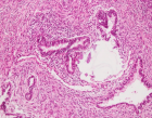
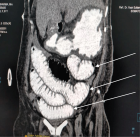
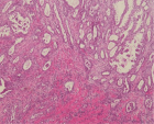
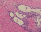
Figure 3:
3: Magnetic resonance imaging (MRI) (a) Sagittal, (b) axial oblique and (c) coronal oblique T2 weighted at T2 show spiculated hypointense areas arranged at confluent angles (white arrows) with loss of cleavage planes between the anterior surface of the sigmoid, the posterior serosa of the uterus and bilateral endometriomas (white arrowheads).
Read Full Article HTML DOI: 10.29328/journal.cjog.1001057 Cite this Article Read Full Article PDF
More Images
Similar Articles
-
Endometriosis as a risk factor for colorectal cancerVíctor Manuel Vargas-Hernández*,José María Tovar- Rodríguez,Víctor Manuel Vargas-Aguilar . Endometriosis as a risk factor for colorectal cancer. . 2020 doi: 10.29328/journal.cjog.1001057; 3: 093-097
-
Epidemiologic aspects and risk factors associated with infertility in women undergoing assisted reproductive technology (ART) in north of IranMarzieh Zamaniyan,Noushin Gordani*,Paniz Bagheri,Kaveh Jafari,Sepideh Peyvandi,Mojtaba Hajihoseini,Robabeh Taheripanah,Siavash Moradi,Salomeh Peyvandi,Arman Alborzi. Epidemiologic aspects and risk factors associated with infertility in women undergoing assisted reproductive technology (ART) in north of Iran. . 2021 doi: 10.29328/journal.cjog.1001079; 4: 015-018
-
Menstruating primary umbilicus cutaneous endometriosis: A case report and review of literatureIdowu Pius Ade-Ojo*,Oluwadare Martins Ipinnimo. Menstruating primary umbilicus cutaneous endometriosis: A case report and review of literature. . 2021 doi: 10.29328/journal.cjog.1001090; 4: 069-071
-
Intravenous leiomyomatosis of the uterus: still discovered on anatomopathological examinationAbir Karoui*,Ahmed Cherif,Olfa Chaffai,Wassim Saidi,Ghada Sahraoui,Sana Menjli,Mohamed Badis Chanoufi,Nadia Boujelbene,Hssine Saber Abouda. Intravenous leiomyomatosis of the uterus: still discovered on anatomopathological examination. . 2022 doi: 10.29328/journal.cjog.1001113; 5: 090-092
Recently Viewed
-
Agriculture High-Quality Development and NutritionZhongsheng Guo*. Agriculture High-Quality Development and Nutrition. Arch Food Nutr Sci. 2024: doi: 10.29328/journal.afns.1001060; 8: 038-040
-
A Low-cost High-throughput Targeted Sequencing for the Accurate Detection of Respiratory Tract PathogenChangyan Ju, Chengbosen Zhou, Zhezhi Deng, Jingwei Gao, Weizhao Jiang, Hanbing Zeng, Haiwei Huang, Yongxiang Duan, David X Deng*. A Low-cost High-throughput Targeted Sequencing for the Accurate Detection of Respiratory Tract Pathogen. Int J Clin Virol. 2024: doi: 10.29328/journal.ijcv.1001056; 8: 001-007
-
A Comparative Study of Metoprolol and Amlodipine on Mortality, Disability and Complication in Acute StrokeJayantee Kalita*,Dhiraj Kumar,Nagendra B Gutti,Sandeep K Gupta,Anadi Mishra,Vivek Singh. A Comparative Study of Metoprolol and Amlodipine on Mortality, Disability and Complication in Acute Stroke. J Neurosci Neurol Disord. 2025: doi: 10.29328/journal.jnnd.1001108; 9: 039-045
-
Development of qualitative GC MS method for simultaneous identification of PM-CCM a modified illicit drugs preparation and its modern-day application in drug-facilitated crimesBhagat Singh*,Satish R Nailkar,Chetansen A Bhadkambekar,Suneel Prajapati,Sukhminder Kaur. Development of qualitative GC MS method for simultaneous identification of PM-CCM a modified illicit drugs preparation and its modern-day application in drug-facilitated crimes. J Forensic Sci Res. 2023: doi: 10.29328/journal.jfsr.1001043; 7: 004-010
-
A Gateway to Metal Resistance: Bacterial Response to Heavy Metal Toxicity in the Biological EnvironmentLoai Aljerf*,Nuha AlMasri. A Gateway to Metal Resistance: Bacterial Response to Heavy Metal Toxicity in the Biological Environment. Ann Adv Chem. 2018: doi: 10.29328/journal.aac.1001012; 2: 032-044
Most Viewed
-
Evaluation of Biostimulants Based on Recovered Protein Hydrolysates from Animal By-products as Plant Growth EnhancersH Pérez-Aguilar*, M Lacruz-Asaro, F Arán-Ais. Evaluation of Biostimulants Based on Recovered Protein Hydrolysates from Animal By-products as Plant Growth Enhancers. J Plant Sci Phytopathol. 2023 doi: 10.29328/journal.jpsp.1001104; 7: 042-047
-
Sinonasal Myxoma Extending into the Orbit in a 4-Year Old: A Case PresentationJulian A Purrinos*, Ramzi Younis. Sinonasal Myxoma Extending into the Orbit in a 4-Year Old: A Case Presentation. Arch Case Rep. 2024 doi: 10.29328/journal.acr.1001099; 8: 075-077
-
Feasibility study of magnetic sensing for detecting single-neuron action potentialsDenis Tonini,Kai Wu,Renata Saha,Jian-Ping Wang*. Feasibility study of magnetic sensing for detecting single-neuron action potentials. Ann Biomed Sci Eng. 2022 doi: 10.29328/journal.abse.1001018; 6: 019-029
-
Pediatric Dysgerminoma: Unveiling a Rare Ovarian TumorFaten Limaiem*, Khalil Saffar, Ahmed Halouani. Pediatric Dysgerminoma: Unveiling a Rare Ovarian Tumor. Arch Case Rep. 2024 doi: 10.29328/journal.acr.1001087; 8: 010-013
-
Physical activity can change the physiological and psychological circumstances during COVID-19 pandemic: A narrative reviewKhashayar Maroufi*. Physical activity can change the physiological and psychological circumstances during COVID-19 pandemic: A narrative review. J Sports Med Ther. 2021 doi: 10.29328/journal.jsmt.1001051; 6: 001-007

HSPI: We're glad you're here. Please click "create a new Query" if you are a new visitor to our website and need further information from us.
If you are already a member of our network and need to keep track of any developments regarding a question you have already submitted, click "take me to my Query."


